43 microscope images with labels
Parts of the Microscope with Labeling (also Free Printouts) Home Microscopes Parts of the Microscope with Labeling (also Free Printouts) By Editorial Team March 7, 2022 A microscope is one of the invaluable tools in the laboratory setting. It is used to observe things that cannot be seen by the naked eye. Table of Contents 1. Eyepiece 2. Body tube/Head 3. Turret/Nose piece 4. Objective lenses 5. LAS X Industry Microscope software for Industry | Products Configure the microscope system with individual user profiles; Make streamlined and flexible measurements when doing analysis ; Enhance your imaging with stitching in X, Y, and Z ; ... area, diameter, angle, or perimeter of objects you mark with adjustable tracing lines, drawing directly in the live images. Add labels for easy analysis.
Microscope Labeled Pictures, Images and Stock Photos Browse 49 microscope labeled stock photos and images available, or start a new search to explore more stock photos and images. Newest results Fluorescent Imaging immunofluorescence of cancer cells growing... Microscope diagram vector illustration. Labeled zoom instrument... Microscope diagram vector illustration.

Microscope images with labels
Microscope Labeling - The Biology Corner The google slides shown below have the same microscope image with the labels for students to copy. I often spend the first day walking students through the steps and having them look at a single slide as we do the steps. Students are often very enthusiastic about using microscopes and will try to start with the high power objective. ZEISS Axioscope 5 Smart Laboratory Microscope With this smart microscope you acquire fluorescent images consisting of up to four different channels. Just push a ... Done. Forget about the 15 steps and clicks to document samples with multiple fluorescent labels. With Smart Microscopy, this is a thing of the past. Axioscope 5 with Axiocam 202 mono and Colibri 3 LED illumination take this ... Labeling the Parts of the Microscope | Microscope World Resources Labeling the Parts of the Microscope This activity has been designed for use in homes and schools. Each microscope layout (both blank and the version with answers) are available as PDF downloads. You can view a more in-depth review of each part of the microscope here. Download the Label the Parts of the Microscope PDF printable version here.
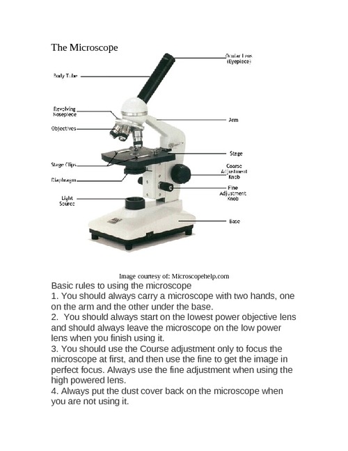
Microscope images with labels. Compound Microscope Parts - Labeled Diagram and their Functions The eyepiece (or ocular lens) is the lens part at the top of a microscope that the viewer looks through. The standard eyepiece has a magnification of 10x. You may exchange with an optional eyepiece ranging from 5x - 30x. [In this figure] The structure inside an eyepiece. The current design of the eyepiece is no longer a single convex lens. Microscope, Microscope Parts, Labeled Diagram, and Functions Revolving Nosepiece or Turret: Turret is the part of the microscope that holds two or multiple objective lenses and helps to rotate objective lenses and also helps to easily change power. Objective Lenses: Three are 3 or 4 objective lenses on a microscope. The objective lenses almost always consist of 4x, 10x, 40x and 100x powers. 18,701 Microscope drawing Images, Stock Photos & Vectors - Shutterstock Find Microscope drawing stock images in HD and millions of other royalty-free stock photos, illustrations and vectors in the Shutterstock collection. Thousands of new, high-quality pictures added every day. Microscope Parts, Function, & Labeled Diagram - slidingmotion Microscope parts labeled diagram gives us all the information about its parts and their position in the microscope. Microscope Parts Labeled Diagram The principle of the Microscope gives you an exact reason to use it. It works on the 3 principles. Magnification Resolving Power Numerical Aperture. Parts of Microscope Head Base Arm Eyepiece Lens
Amazon.com: Magnifying Glasses 8X 15X 23X Magnifier LED … About this item . Double eye magnifying glasses magnifier loupe, with 2pcs adjustable LEDs to help it work in low-light conditions ; Left right double eye patches magnifierloupe with adjustable LED to help work in low-light conditions.Set of 2 magnifying glasses mounted on a one-size-fits-all eyeglass frame for easy hands-free operation. Parts of a Simple Microscope - Labeled (with diagrams) image 2: A simple microscope commonly used by students for studying minute objects. image source: imimg.com. picture 3: It is the latest design of a simple microscope - advanced features than the conventional simple microscopes. ... image 5: A modern simple microscope with the different parts labeled. image source: laboratoryinfo.com. The ... ZEISS Elyra 7 with Lattice SIM² Super-Resolution Microscope Images of Cos-7 cell stained with anti-alpha-Tubulin Alexa fluor 488 were processed with the conventional SIM algorithms based on generalized Wiener filter and with the novel SIM² reconstruction. The images show an improvement of resolution for SIM² compared to SIM. Objective: Plan-Apochromat 63× / 1.4 Oil. PDF Label parts of the Microscope Label parts of the Microscope: . Created Date: 20150715115425Z
300+ Free Microscope & Laboratory Images - Pixabay 399 Free images of Microscope Related Images: laboratory science bacteria research scientist lab biology chemistry medical Find your perfect microscope image. Free pictures to download and use in your next project. Mitosis Images Labeled | Virtual Anatomy Lab VAL - ncccval Endocrine Rabbit Dissection Unlabeled. Cardiovascular. Cardiovascular Histology Labeled. Cardiovascular Histology Unlabeled. Cardiovascular Models Labeled. Cardiovascular Models Unlabeled. Cardiovascular Sheep Heart Dissect-L. Cardiovascular Sheep Heart Disect-U. Cardiovascular Cat Dissection Labeled. Microscope Label Interactive Worksheets & Teaching Resources | TpT 12. $1.89. PDF. Students will complete a timeline of the history of the microscope, label a diagram, and create a pocket foldable with terms and definition cards. The timeline can be completed according to the teacher's directions or like the answer key example. Optional cut & paste images and a QR code are a. Microscope Types (with labeled diagrams) and Functions The working principle of a simple microscope is that when a lens is held close to the eye, a virtual, magnified and erect image of a specimen is formed at the least possible distance from which a human eye can discern objects clearly. Simple microscope labeled diagram Simple microscope functions It is used in industrial applications like:
33 Label Of Compound Microscope - Labels Database 2020
Compound Microscope Parts, Functions, and Labeled Diagram Compound Microscope Definitions for Labels Eyepiece (ocular lens) with or without Pointer: The part that is looked through at the top of the compound microscope. Eyepieces typically have a magnification between 5x & 30x. Monocular or Binocular Head: Structural support that holds & connects the eyepieces to the objective lenses.
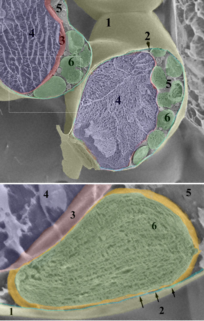
Leaf chloroplast
Explanation and Labelled Images - New York Microscope Company Another use of fluorescence imaging is Fluorescence Speckle Microscopy. It is a technology that uses fluorescence labeled macromolecular assemblies such as cytoskeletal protein to study movement and turnover rates. Fluorescence microscopy staining also is helpful in the field of mineralogical applications. It is routinely used for the study of ...
Post a Comment for "43 microscope images with labels"