40 microscope with labels and functions
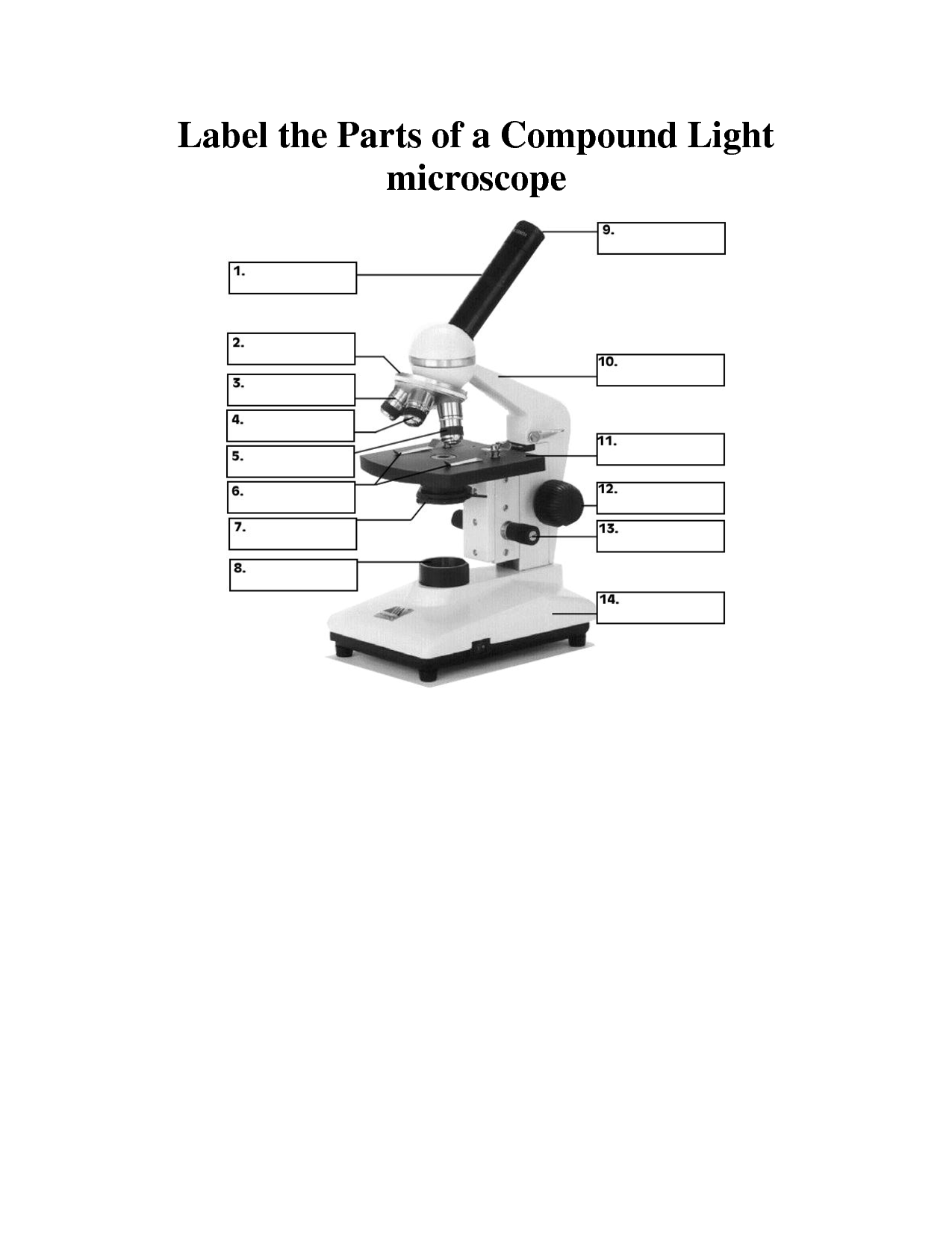
PDF Parts of a Microscope Printables - Homeschool Creations Label the parts of the microscope. You can use the word bank below to fill in the blanks or cut and paste the words at the bottom. Microscope Created by Jolanthe @ HomeschoolCreations.net. Parts of a eyepiece arm stageclips nosepiece focusing knobs illuminator stage objective lenses Virtual microscope tutorial - palada.me Jul 31, 2022 · 2021 There are a couple virtual microscope labs available online you can have If you would like a free powerpoint I created for this lesson, MyScope™ is a fantastic online training tool for microscopy and microanalysis. The Paper Microscope. Therefore animal cells, plant cells, protozoa, bacteria can be [email protected]
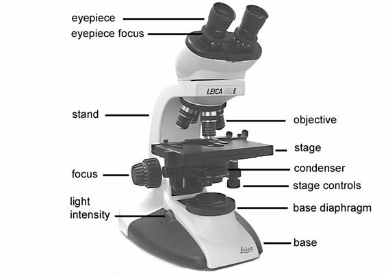
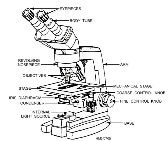
Microscope Parts and Functions First, the purpose of a microscope is to magnify a small object or to magnify the fine details of a larger object in order to examine minute specimens that cannot be seen by the naked eye. Here are the important compound microscope parts... Eyepiece: The lens the viewer looks through to see the specimen.

Microscope with labels and functions
Microscope Diagram and Functions | Science fair projects, Microscope ... A Study of the Microscope and its Functions With a Labeled Diagram. To better understand the structure and function of a microscope, we need to take a look at the labeled microscope diagrams of the compound and electron microscope. These diagrams clearly explain the functioning of the microscopes along with their respective parts. Parts of a Microscope with Their Functions - Microbe Online Aug 2, 2022 — Microscope is a piece of laboratory optical equipment that is used to magnify small objects. The compound microscope has many parts. The Parts of a Microscope (Labeled) Printable - TeacherVision The Parts of a Microscope (Labeled) Printable. Download. Add to Favorites. Share. This diagram labels and explains the function of each part of a microscope. Use this printable as a handout or transparency to help prepare students for working with laboratory equipment.
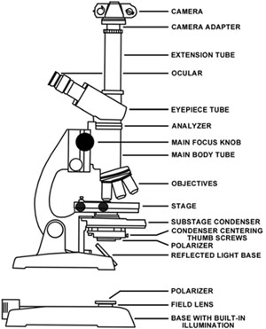
Microscope with labels and functions. Microscope Parts & Functions - AmScope Microscope Parts and Functions Invented by a Dutch spectacle maker in the late 16th century, compound light microscopes use two sets of lenses to magnify images for study and observation. The first set of lenses are the oculars, or eyepieces, that the viewer looks into; the second set of lenses are the objectives, which are closest to the specimen. Microscope With Labeled Parts and Functions - 24 Hours Of Biology Optical parts and the functions The optical parts of the microscope are used to view, enlarge, and produce an image from a sample placed on a slide. These parts include Eyepiece: Eyepiece also contains ocular lens. It enhance the image of the viewer. This part is used for checking through the microscope. Eyepiece is found at the upper part of it. Parts of a microscope with functions and labeled diagram - Microbe Notes Microscopes are instruments that are used in science laboratories to visualize very minute objects such as cells, and microorganisms, giving a contrasting image that is magnified. Microscopes are made up of lenses for magnification, each with its own magnification powers. Simple Microscope - Parts, Functions, Diagram and Labelling Parts of the optical parts are as follows: Mirror - A simple microscope has a plano-convex mirror and its primary function is to focus the surrounding light on the object being examined. Lens - The biconvex lens is placed above the stage and its function is to magnify the size of the object being examined.
Compound Microscope- Definition, Labeled Diagram, Principle, Parts, Uses A compound microscope is of great use in pathology labs so as to identify diseases. Various crime cases are detected and solved by drawing out human cells and examining them under the microscope in forensic laboratories. The presence or absence of minerals and the presence of metals can be identified using compound microscopes. Histology - Yale University Bone is a tissue in which the extracellular matrix has been hardened to accommodate a supporting function. The fundamental components of bone, like all connective tissues, are cells and matrix. There are three key cells of bone tissue. They each have unique functions and are derived from two different cell lines. Microscope labeling and functions Flashcards | Quizlet Microscope labeling and functions STUDY Flashcards Learn Write Spell Test PLAY Match Gravity Created by mveet Terms in this set (27) Separates the eyepiece lens from the objective lenses Body Tube Holds the low-power and high-power objective lenses; allows the lenses to rotate for viewing Revolving Nosepiece Magnifies about 4x Compound Microscope: Definition, Diagram, Parts, Uses, Working ... - BYJUS The compound microscope is mainly used for studying the structural details of cell, tissue, or sections of organs. The parts of a compound microscope can be classified into two: Non-optical parts Optical parts Non-optical parts Base The base is also known as the foot which is either U or horseshoe-shaped.
Microscope Parts, Function, & Labeled Diagram - slidingmotion Microscope parts labeled diagram gives us all the information about its parts and their position in the microscope. Microscope Parts Labeled Diagram The principle of the Microscope gives you an exact reason to use it. It works on the 3 principles. Magnification Resolving Power Numerical Aperture. Parts of Microscope Head Base Arm Eyepiece Lens Microscope Quiz: How Much You Know About Microscope Parts And Functions ... Projects light upwards through the diaphragm, the specimen, and the lenses. 5. Is used to regulates the amount of light on the specimen. Supports the slide being viewed. Moves the stage up and down for focusing. 6. Is used to support the microscope when carried. Moves the stage slightly to sharpen the image. Parts of the Microscope with Labeling (also Free Printouts) Let us take a look at the different parts of microscopes and their respective functions. 1. Eyepiece it is the topmost part of the microscope. Through the eyepiece, you can visualize the object being studied. Its magnification capacity ranges between 10 and 15 times. 2. Body tube/Head It is the structure that connects the eyepiece to the lenses. A Study of the Microscope and its Functions With a Labeled Diagram ... A Study of the Microscope and its Functions With a Labeled Diagram To better understand the structure and function of a microscope, we need to take a look at the labeled microscope diagrams of the compound and electron microscope. These diagrams clearly explain the functioning of the microscopes along with their respective parts.
Microscope With Labels Clip Art at Clker.com - vector clip art online, royalty free & public domain
ZEISS Axioscan 7 Microscope Slide Scanner Digitize your specimens with Axioscan 7 – the reliable, reproducible way to create high-quality virtual microscope slides. Axioscan 7 combines qualities that you would not expect to get in a slide scanner: high speed digitization and outstanding image quality plus an unrivaled variety of imaging modes are all available in a fully automated and easy to operate system.
8 Best Images of Lens Diagram Worksheet - Microscope with Labeled Parts, Label Eye Parts ...
Stereo Microscope Parts A stereo microscope has three key parts: Viewing Head/Body that houses the optical components in the upper part of the microscope. Focus Block that attaches the microscope head to the stand and focuses the microscope. Stand that supports the microscope and houses any integrated illumination. Stereo microscopes are increasingly modular.
8 Best Images of Lens Diagram Worksheet - Microscope with Labeled Parts, Label Eye Parts ...
Parts of a Microscope Labeling Activity - Storyboard That Create a poster that labels the parts of a microscope and includes descriptions of what each part does. Click "Start Assignment". Use a landscape poster layout (large or small). Search for a diagram of a microscope. Using arrows and textables label each part of the microscope and describe its function.

34 Label Parts Of A Microscope - Labels 2021
Microscope: Parts Of A Microscope With Functions And Labeled Diagram. Q. Define a Microscope. Ans. Microscopes are instruments that are used in science laboratories, to visualize very minute objects such as cells, and microorganisms, giving a contrasting image, that is magnified. Q. State functions of a microscope. Ans. A microscope is usually used for the study of microscopic algae, fungi, and biological specimens.

7th Grade Science Class: Cells and "E" Lab
Parts of Stereo Microscope (Dissecting microscope) - labeled diagram ... Stereo microscopes (also called Dissecting microscope) are branched out from other light microscopes for the application of viewing "3D" objects. These include substantial specimens, such as insects, feathers, leaves, rocks, sand grains, gems, coins, and stamps, etc. Functionally, a stereo microscope is like a powerful magnifying glass.

Microscope World Blog: Cerebellum under the Microscope
Microscope Parts, Functions, and Labeling Flashcards | Quizlet Start studying Microscope Parts, Functions, and Labeling. Learn vocabulary, terms, and more with flashcards, games, and other study tools.
3.3: Pre-lab Questions - Biology LibreTexts
Confocal Microscopy - an overview | ScienceDirect Topics A confocal microscope was invented in 1951 by Marvin Minsky, a postdoctoral fellow at Harvard University studying neural networks in living brain (Minsky, 1988).In 1957, Minsky patented the concept of confocal imaging, the illumination and detection of a single diffraction-limited spot in a specimen (Fig. 1A).
Labels Of The Microscope
Parts of a Compound Microscope and Their Functions - NotesHippo Compound microscope uses in forensic labs it easy to detect human fingerprints. A compound microscope can be used to detect the presence of metals. The use of a compound microscope makes studying germs and viruses much easier. Compound microscope uses in schools makes learning biology easy for all children.

Label diagram of compound microscope - Science - The Fundamental Unit of Life - 12499729 ...
Labeling the Parts of the Microscope | Microscope World Resources Labeling the Parts of the Microscope. This activity has been designed for use in homes and schools. Each microscope layout (both blank and the version with answers) are available as PDF downloads. You can view a more in-depth review of each part of the microscope here.

labeled microscope for kids - Google Search | {School} - Science | Biology for kids, Teaching ...
22 Parts Of a Microscope With Their Function And Labeled Diagram A light microscope is a type of microscope that commonly uses visible light and a system of lenses to generate magnified images of small objects whereas electron microscope is a microscope that uses a beam of accelerated electrons as a source of illumination. It is a special type of microscope with a high resolution of images.
Label Compound Microscope: SP17C16 BIO2921VB30 Microbiology Laboratory
Compound Microscope Parts, Functions, and Labeled Diagram Compound Microscope Parts, Functions, and Labeled Diagram Parts of a Compound Microscope Each part of the compound microscope serves its own unique function, with each being important to the function of the scope as a whole.

Instructions - Microbiology Action - 78 Steps Health
Microscope, Microscope Parts, Labeled Diagram, and Functions Illuminator: Illuminator is the most important microscope parts and it serve as light source for a microscope during slide specimen visualization. It is a continuous source of light (110 volts) used in place of a mirror. The mirror of microscope is used to reflect light from the external light source up through the bottom of the stage.
Compound Microscope Diagram With Labels - Micropedia
LAS X Industry Microscope software for Industry | Products ... Measure parameters, such as the length, area, diameter, angle, or perimeter of objects you mark with adjustable tracing lines, drawing directly in the live images. Add labels for easy analysis. Apply measurements to several images to determine statistical trend and compare data in measurement templates.
Molecular Expressions: Microscopy Publications - Photomicrography in the Geological Sciences
Electron microscope - Wikipedia An electron microscope is a microscope that uses a beam of accelerated electrons as a source of illumination. As the wavelength of an electron can be up to 100,000 times shorter than that of visible light photons , electron microscopes have a higher resolving power than light microscopes and can reveal the structure of smaller objects.

30 Label The Indicated Parts Of The Microscope - Label Ideas 2020
Compound Microscope Parts - Labeled Diagram and their Functions Two adjustment knobs are used to focus the microscope: fine focus knob and coarse focus knob. Both knobs can move the stage up and down. You should use the coarse focus knob to bring the specimen into approximate or near focus. Then you use the fine focus knob to sharpen the focus quality of the image.

Anatomy and Physiology I Coursework: Connective Tissues
Microscopy- History, Classification, Terms, Diagram - The Biology Notes Fluorescence Microscopy is a microscopy technique that uses a fluorescent microscope with a UV light source. It is widely used in detecting antigens, antibodies, and other macromolecules. Fluorescence Microscope 5. Confocal Microscopy Confocal Microscopy is a newer microscopy technique that uses a focused laser beam.

33 Microscope Diagram To Label - Labels Database 2020
Label the microscope — Science Learning Hub Jun 08, 2018 · All microscopes share features in common. In this interactive, you can label the different parts of a microscope. Use this with the Microscope parts activity to help students identify and label the main parts of a microscope and then describe their functions. Drag and drop the text labels onto the microscope diagram. If you want to redo an ...
Post a Comment for "40 microscope with labels and functions"