39 brain mri with labels
Cross-sectional anatomy of the brain - e-Anatomy - IMAIOS We created a brain atlas that is an interactive tool for studying the conventional anatomy of the normal brain based on a magnetic resonance imaging exam of the axial brain. Anatomical structures and specific areas are visible as interactive labeled images. Cross sectional anatomy: MRI of the brain. An MRI was performed on a healthy subject ... New MRI probe can reveal more of the brain's inner workings Traditional fMRI imaging measures changes to blood flow in the brain, as a proxy for neural activity. When neurons receive signals from other neurons, it triggers an influx of calcium, which causes...
New MRI probe can reveal more of the brain's inner workings Using this technique, which involves genetically targeting the MRI probe to specific populations of cells in animal models, the researchers were able to identify neural populations involved in a circuit that responds to rewarding stimuli. The new MRI probe could also enable studies of many other brain circuits, the researchers say.

Brain mri with labels
MRI sequences (overview) | Radiology Reference Article - Radiopaedia This leads to a broad categorization as follows: T1 weighted (T1W) gadolinium enhanced fat suppressed T2 weighted (T2W) fat suppressed fluid attenuated susceptibility sensitive proton density (PD) fat suppressed diffusion weighted flow sensitive MR angiography (MRA) MR venography (MRV) CSF flow studies miscellaneous Segmentation Labels and Radiomic Features for the Pre-operative Scans ... this data container describes both computer-aided and manually-corrected segmentation labels for the pre-operative multi-institutional scans of the cancer genome atlas (tcga) low grade glioma (lgg) collection, publicly available in the cancer imaging archive (tcia), coupled with a rich panel of radiomic features along with their corresponding … Tutorials/SegBrainSuite - Brainstorm You can use the free BrainSuite and SVReg software package to extract segmented brain surfaces from a T1-weighted MRI image. ... Checking the box next to "Register and label brain" when running BrainSuite's Cortical Surface Extraction sequence causes BrainSuite to run a companion program called SVReg that implements an automatic parcellation of ...
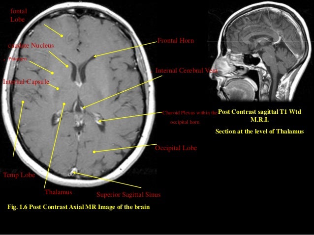
Brain mri with labels. Brain MRI: How to read MRI brain scan | Kenhub MRI is the most sensitive imaging method when it comes to examining the structure of the brain and spinal cord. It works by exciting the tissue hydrogen protons, which in turn emit electromagnetic signals back to the MRI machine. The MRI machine detects their intensity and translates it into a gray-scale MRI image. Anatomical diagrams of the brain - e-Anatomy - IMAIOS The anatomical study of the brain continues with the study of commissural fibres, including the corpus callosum, the fornix, septum pellucidum, the anterior commissure and then through the study of the basal ganglia (lenticular nucleus, nucleus caudate and globus pallidus), related structures (medial and lateral blade cords, internal and ... Epigenetic MRI: Noninvasive imaging of DNA methylation in the brain The 13 C-NMR detects labeled DNA and age-dependent labeling in the brain. ( A) Sample setup for spectroscopy and imaging experiments. The intact brain hemisphere was submerged in PFC (perfluorocarbon) oil for susceptibility matching to improve magnetic field homogeneity. ( B) A 3D MR image from one of the brain samples. Can anyone suggest an online tool for entering ... - ResearchGate In recent years several novel approaches for constructing MRI based brain parcellations have been developed with promising results. In the absence of ground truth, several evaluation approaches ...
Brain Tumor MRI Dataset | Kaggle Br35H This dataset contains 7022 images of human brain MRI images which are classified into 4 classes: glioma - meningioma - no tumor and pituitary. no tumor class images were taken from the Br35H dataset. MRI study guide: Quizzes, test questions & flashcards - Kenhub The concept is simple. Click below to download the unlabeled MRI test questions worksheet, and label the name of the structure you see in each MRI image. Once you've completed that, you can also download the labeled version of the worksheet to find out how your MRI questions and answers match up, and to make some notes. Labeled imaging anatomy cases | Radiology Reference Article ... This article lists a series of labeled imaging anatomy cases by body region and modality. Brain CT head: non-contrast axial CT head: non-contrast coronal CT head: non-contrast sagittal CT head: angiogram axial CT head: angiogram coronal CT... ASLPrep: a platform for processing of arterial spin labeled MRI and ... Arterial spin labeled (ASL) magnetic resonance imaging (MRI) is the primary method for noninvasively measuring regional brain perfusion in humans. We introduce ASLPrep, a suite of software ...
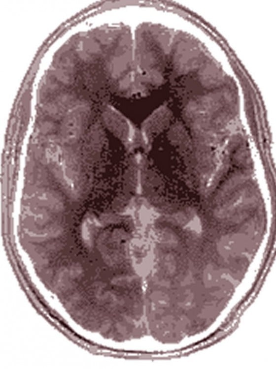
Normal brain imaging examples | Radiology Reference Article ... Citation, DOI & article data. This article lists examples of normal imaging of the brain and surrounding structures, divided by modality and protocol. CT. CT (routine) example 1: C- axial, coronal, sagittal. example 2: C- axial, coronal, sagittal & axial bone. example 3: C- axial, C+ axial, coronal, sagittal. example 4: C- axial, coronal. MRI Images Epigenetics in the Brain - Neuroscience News Getting this label into the brain is easy and does no harm to the body. We'll give it to people through the diet and then we can detect the signal." Their first application of the approach will likely occur in studies comparing the brains of people with and without neurodegenerative disease, he said. About this neuroimaging research news Automated segmentation of multiparametric magnetic resonance ... - Nature A hybrid semi-automated and manual approach was used to label MRI/MRAs with arteries, veins, brain parenchyma, cerebral spinal fluid (CSF), and embolized vessels. Next, these labels were used to ... Ventricles of the brain: Anatomy and pathology | Kenhub The fluid (cerebrospinal fluid) is produced in the ventricular system of the brain. There are four such hollow spaces in the brain that house cerebrospinal fluid (CSF): two lateral ventricles, a third ventricle and a fourth ventricle. This article will look at the structure of this system and how it helps the brain. Contents Choroid plexus
MRI Brain Planning
Anatomy of the face and neck (MRI) - e-Anatomy - IMAIOS The bones of the face and neck were labeled using different colors to facilitate comprehension. The bone structures are rather more difficult to view on a weighted MRI T2 than on a CT-Scan: for more details on the bones of the face, please refer to the e-Anatomy module "Face-CT-Scan". The teeth were numbered using the FDI World Dental ...
MRI SECTIONAL ANATOMY OF BRAIN
Arterial spin labeling MR perfusion - Radiopaedia Arterial spin labeling (ASL) MR perfusion is an MR perfusion technique which does not require intravenous administration of contrast (unlike DSC perfusion and DCE perfusion ). Instead it exploits the ability of MRI to magnetically label arterial blood below the imaging slab. The parameter most commonly derived is cerebral blood flow (CBF).

UMD PSYC E-News: RA positions focusing on functional MRI and Autism Spectrum Disorder!
Arterial Spin Labeling Imaging Assessment of Cerebrovascular Reactivity ... Each participant underwent a brain MRI study, and CVR was calculated as the cerebral blood flow (CBF) reduction using arterial spin labeling (ASL) between baseline and 10 min after an intravenous dipyridamole injection (0.57 mg/kg).

February | 2011 | Thought Broadcast
brain midsagittal view labels - Sagittal Section Of Brain Labeled | mri ... Brain Midsagittal View Labels images that posted in this website was uploaded by Media.nbcmontana.com. Brain Midsagittal View Labels equipped with a HD resolution 1064 x 851.You can save Brain...

Radiology MRI: Neonatal Intraventricular Hemorrhage
Classification of brain tumours in MR images using deep ... - Nature A brain tumour is the growth of abnormal cells in the brain. Brain tumours are classified based on their speed of growth and the likeness of them growing back after treatment. They are mainly...
Brain Anatomy Differences Between Autistic and Typically Developing Individuals Are ...
Artificial Intelligence-based MRI Images for Brain in Prediction of ... 3.1. Brain MRI features of AD patients. Figure 3 below showed MRI images of the brain of AD patients and normal people. In the MRI images of the brain of AD patients, brain atrophy was observed, which mainly occurred in the hippocampus, parahippocampal gyrus, and medial temporal lobes. At the same time, ventricles were dilated.
A Hybrid Approach for Automatic Classification of Brain MRI Using Genetic Algorithm and Support ...
New machine learning model flags abnormal brain scans in real-time "Having previously built and validated a labeled head MRI dataset using cutting edge machine learning methodology through a team of data scientists and hospital radiologists, the same team have now...
MRI Procedure of Brain
Cerebral angiography - e-Anatomy - IMAIOS IMAIOS and selected third parties, use cookies or similar technologies, in particular for audience measurement. Cookies allow us to analyze and store information such as the characteristics of your device as well as certain personal data (e.g., IP addresses, navigation, usage or geolocation data, unique identifiers).
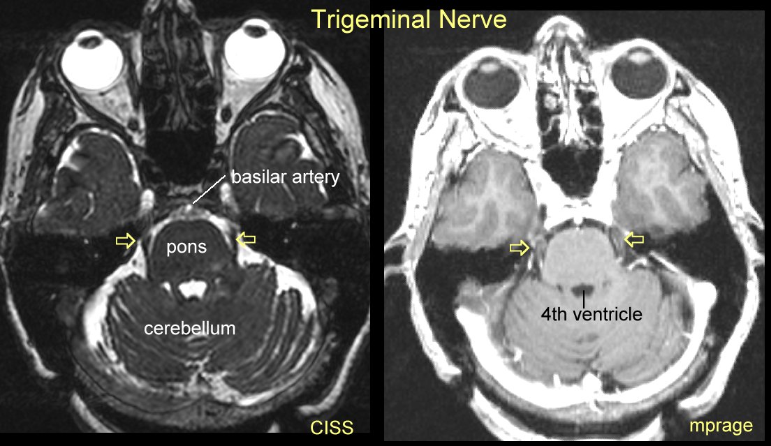
Gamma Knife for Trigeminal Neuralgia
MR cerebral venography | Radiology Reference Article - Radiopaedia The purpose of this exam is to visualize the cerebral veins and venous sinuses allowing their anatomy and patency to be assessed either using non-contrast or contrast-enhanced depiction of venous blood flow. Technique Multiple MRI techniques can be used to visualize the cerebral veins 1 . Non-contrast-enhanced flow related MRI

Neuroanatomy - encyclopedia article - Citizendium
Brain: Atlas of human anatomy with MRI - e-Anatomy - IMAIOS MRI Atlas of the Brain. This page presents a comprehensive series of labeled axial, sagittal and coronal images from a normal human brain magnetic resonance imaging exam. This MRI brain cross-sectional anatomy tool serves as a reference atlas to guide radiologists and researchers in the accurate identification of the brain structures.

Radiodiagnosis - Imaging is Amazing-Interesting cases: Phenytoin associated Cerebellar atrophy - MRI
Tutorials/LabelFreeSurfer - Brainstorm Authors: Francois Tadel. The open-source software FreeSurfer can be used to extract the cortical envelope from a T1/T2 MRI and register it to an atlas. The process is fully automatic and the results can be imported in Brainstorm with just a few mouse clicks. If you are using FreeSurfer, please register on their website ( registration page) and ...

Gamma Knife for Trigeminal Neuralgia
Tutorials/SegBrainSuite - Brainstorm You can use the free BrainSuite and SVReg software package to extract segmented brain surfaces from a T1-weighted MRI image. ... Checking the box next to "Register and label brain" when running BrainSuite's Cortical Surface Extraction sequence causes BrainSuite to run a companion program called SVReg that implements an automatic parcellation of ...

Medical Legal Demonstrative Evidence
Segmentation Labels and Radiomic Features for the Pre-operative Scans ... this data container describes both computer-aided and manually-corrected segmentation labels for the pre-operative multi-institutional scans of the cancer genome atlas (tcga) low grade glioma (lgg) collection, publicly available in the cancer imaging archive (tcia), coupled with a rich panel of radiomic features along with their corresponding …

Mouse brain seen in sharpest detail ever | Kurzweil
MRI sequences (overview) | Radiology Reference Article - Radiopaedia This leads to a broad categorization as follows: T1 weighted (T1W) gadolinium enhanced fat suppressed T2 weighted (T2W) fat suppressed fluid attenuated susceptibility sensitive proton density (PD) fat suppressed diffusion weighted flow sensitive MR angiography (MRA) MR venography (MRV) CSF flow studies miscellaneous

MRI of Brain - Stock Image - M134/0816 - Science Photo Library

MRI Brain Poster | Zazzle.com

Radiology MRI: Occipital Infarct
Post a Comment for "39 brain mri with labels"