38 colon diagram with labels
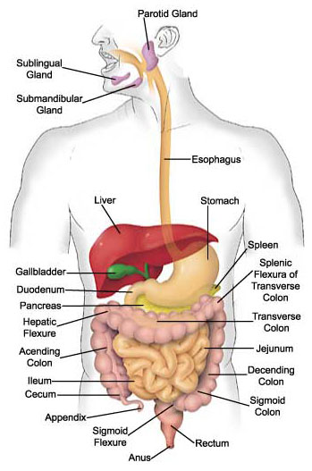
Colon (Large Intestine): Anatomy, Function, Structure Sigmoid colon: The S-shaped connection between the last part of the colon and the rectum, located on the bottom left side of the abdomen is called the sigmoid colon. 2 Size and Length This organ is called the large intestine because of the diameter (width) of the intestine; it is much wider than the small intestine, but also much shorter. Colon Anatomy (with Small Intestine Label) - NCI Visuals Online 720x602. View. Download. Title: Colon Anatomy (with Small Intestine Label) Description: Drawing shows the cecum, ascending colon, transverse colon, descending colon, sigmoid colon, rectum, and anal canal. Also shown is the small intestine. The cecum connects the small intestine to the colon.
Colonoscopy Measurements (cm) from Anal Verge | SEER Training Types of Surgery: Colon; Types of Surgery: Rectum; Radiation Therapy; Commonly Used Drugs; For hands-on exercises, please go to SEER*Educate. Resources. Archived Modules. Updates. Acknowledgements. Colonoscopy Measurements (cm) from Anal Verge. Return to Anatomy of Colon and Rectum. Follow SEER. Contact Information.

Colon diagram with labels
endocrine system diagram to label Digestive System With Labels Focusing On The Colon, Rectum, And Anus . ... Label The Human Stomach On A Diagram The Four Main Regions Of Stomach medicinebtg.com. stomach label anatomy diagram digestive human sphincter regions system labeled identify physiology main curvatures four its google body models types. support.microsoft.com › en-us › officeUse the Create Diagram from Data wizard - support.microsoft.com You can use the Create Diagram from Data wizard to create a detailed, polished Visio flowchart from an Excel workbook. Follow the steps in the wizard and use this help information if you have questions in each step. For more information about Data Visualizer, see Create a Data Visualizer diagram. diagram of female pelvis Transvaginal sagittal plane sonography anatomy pelvis diagram female normal scan uterus sonogram corpus showing figure. Pelvic floor female 3d. ... And Pelvis Labeled Structures Include Large Bowel Colon Diagrams Female medicinebtg.com. colon pelvis bowel medicinebtg urinary excretory. CT Abdomen And Pelvis With IV Contrast. Left Panel ...
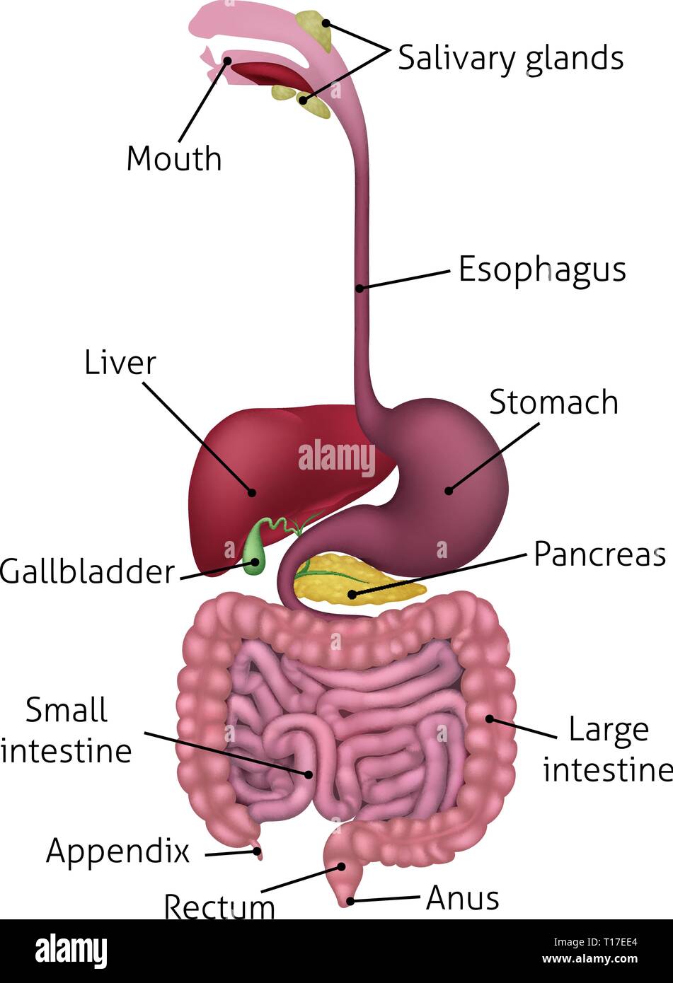
Colon diagram with labels. PDF ANATOMIC DRAWINGS OF THE DIGESTIVE SYSTEM Esophageal sphincter Liver ... Colon (C18._).0 Cecum.1 Appendix.2 Ascending.3 Hepatic flex..4 Transverse.5 Splenic flex..6 Descending.7 Sigmoid.8 Overlapping.9 Colon, NOS Yes Yes Yes Yes Yes Yes Yes Yes Yes Yes Yes Yes Yes Yes Yes Yes Yes Yes Yes Yes Yes Yes Yes Yes Yes Yes Yes Yes Yes Yes Yes Yes Yes Yes Yes Yes Yes Yes Yes Yes Yes Yes Yes Yes Yes Yes Yes Yes Yes Yes Yes ... Digestive System with Labels Focusing on the Colon, Rectum, and Anus Black and white illustration of the digestive system with colon, rectum, and anus highlighted and parts labeled: esophagus, stomach, liver, gallbladder, duodenum ... › nutritionsource › healthyHealthy Eating Plate vs. USDA’s MyPlate | The Nutrition ... The Healthy Eating Plate encourages consumers to choose fish, poultry, beans or nuts, protein sources that contain other healthful nutrients. It encourages them to limit red meat and avoid processed meat, since eating even small quantities of these foods on a regular basis raises the risk of heart disease, diabetes, colon cancer, and weight gain. Sigmoid colon - Definition, Anatomy and Function | Kenhub Sigmoid colon - ventral view The gastrointestinal system is divided into the foregut, midgut and hindgut. The foregut stretches from the oesophagus to the major duodenal papilla, the midgut from the major duodenal papilla to two thirds of the transverse colon, and the hindgut from this point to the pectinate line of the rectum. Neurovasculature
Cecum Histology Slide with Labeled Image and Diagram This taenia coli is not commonly found in all species. If you want to know more about the histological features of the colon, you may read the following article. Histological features of the colon with the labeled diagram and microscope image; You will learn the details histological features of the colon and compare it with the cecum microscope ... Digestive tract - labeled | Media Asset | NIDDK The lower GI tract consists of the large intestine—which includes the colon and rectum—and anus. Diseases or Conditions. Digestive Diseases. File Size. 420 KB | 1386 x 1698. File Type. JPG. Related Keywords English labels anatomy anus rectum colon small intestine duodenum stomach esophagus mouth digestive tract. Share this page. Print ... Intestines (Anatomy): Picture, Function, Location, Conditions Velvety tissue lines the small intestine, which is divided into the duodenum, jejunum, and ileum. The large intestine (colon or large bowel) is about 5 feet long and about 3 inches in diameter. The... Histology | Colon The 4 basic layers of the colon: This diagram illustrates the 4 basic layers of the colon. The inner pink layer is the mucosa, the yellow layer beneath the mucosa is called the submucosa, while the red layer is the muscular layer (muscularis) and the 4 th. layer is called the serosa or adventitia.. Courtesy Ashley Davidoff MD
How does the bowel work? Bowel information with diagrams - Macmillan ... The small bowel is part of the digestive system. It is between the stomach and the large bowel (colon). The small bowel is between 4 and 6 metres long. It folds many times to fit inside the tummy (abdomen). It breaks down food, allowing vitamins, minerals and nutrients to be absorbed into the body. The small bowel is made up of three main parts: Body Cavities and Membranes: Labeled Diagram, Definitions The 3 meningeal layers are labeled with the stars. The outermost layer of the meninges is the dura mater, which is located beneath the skull. Below the dura mater is the arachnoid, which is the middle meningeal layer. There is a space below the arachnoid called the subarachnoid space, and this is where the CSF is located. 40 Colon diagram Vector Images, Colon diagram Illustrations - Depositphotos 40 Colon diagram Stock Vector Images, Royalty-free Colon diagram Drawings & Illustrations. VectorMine Crohns disease vector illustration. Labeled diagram with diagnosis. VectorMine Ulcerative colitis vector illustration. Labeled anatomical infographic. en.wikipedia.org › wiki › Probability_distributionProbability distribution - Wikipedia A probability distribution is a mathematical description of the probabilities of events, subsets of the sample space.The sample space, often denoted by , is the set of all possible outcomes of a random phenomenon being observed; it may be any set: a set of real numbers, a set of vectors, a set of arbitrary non-numerical values, etc.
Colon Anatomy - Human Body Diagrams - Medical Art Library The large intestine begins at the cecum. The ileum (small intestine) ends where it connects to the cecum at the ileocecal junction. The colon is divided into four parts: the ascending, transverse, descending and sigmoid. The ascending and transverse colon meet at the right hepatic flexure (near the liver). The transverse and descending colon ...
› stock-photo › female-anatomy-diagramFemale Anatomy Diagram Stock Photos and Images - Alamy Find the perfect female anatomy diagram stock photo. Huge collection, amazing choice, 100+ million high quality, affordable RF and RM images. No need to register, buy now!
verificationguide.com › systemverilog › systemveriAssertions in SystemVerilog Immediate and Concurrent ... The optional statement label (identifier and colon) creates a named block around the assertion statement; The action block is executed immediately after the evaluation of the assert expression; The action_block specifies what actions are taken upon success or failure of the assertion; action_block; pass_statement; else fail_statement;
Label Digestive System Diagram Printout - EnchantedLearning.com Read the definitions below, then label the digestive system anatomy diagram. anus - the opening at the end of the digestive system from which feces (waste) exits the body. appendix - a small sac located on the cecum. ascending colon - the part of the large intestine that run upwards; it is located after the cecum.


Post a Comment for "38 colon diagram with labels"